62 The Basics of Brain Structure and Function
Alexandria Lewis
Left vs. Right Brain Myth
overview
Some of the following content is adapted from Human Biology Pressbooks by Christine Miller (Creative Commons Attribution Noncommercial)
The adult brain makes up around 2% of the body’s weight, and it uses about 20% of the body’s total energy. The brain contains an estimated 100 billion neurons, and each neuron has thousands of synaptic connections to other neurons. The brain controls such mental processes as reasoning, imagination, memory, and language. It also interprets information from the senses and commands the body to respond appropriately. It controls basic physical processes (such as breathing and heartbeat), as well as voluntary activities (such as walking and writing).
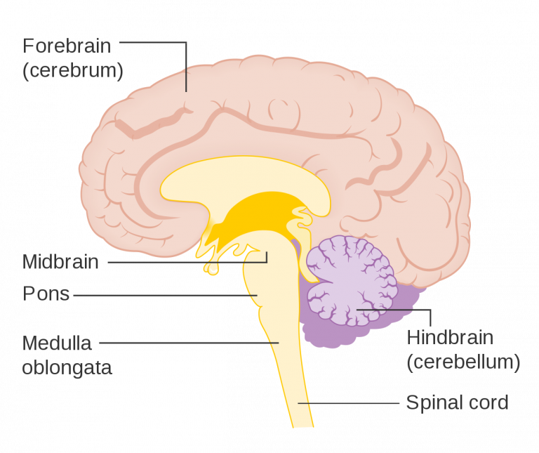
Hindbrain
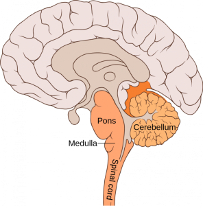
The hindbrain includes the cerebellum, medulla oblongata, and the pons; it is the lowest part of the brain. The hindbrain resembles a stalk and platform on which the cerebrum is perched. The components of the hindbrain connect the rest of the brain with the spinal cord and passes nerve impulses between the brain and spinal cord.
Note: Click on a heading to expand the information.
Midbrain
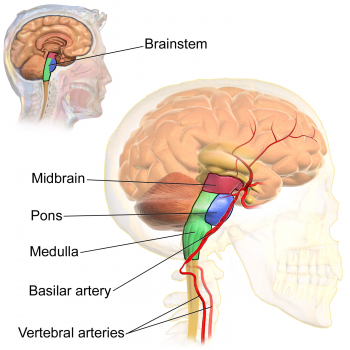
The reticular activating system (RAS) is responsible for the sleep-wake cycle and wakefulness. The RAS also regulates attention, ability to focus and arousal.
Forebrain
The forebrain is the anterior (forwardmost) part of the brain and includes the cerebrum, the thalamus and hypothalamus, hippocampus, amygdala, and limbic system. This portion of the brain is responsible for processing incoming sensory information, performing complex cognitive activities (speech, abstract thought, etc.) and governing voluntary motor movements. The forebrain also controls body temperature, reproductive functions, eating, sleeping and the display of emotions.
Cerebrum vs. Cerebral cortex
| Feature | Cerebrum | Cerebral Cortex |
|---|---|---|
| Definition | Largest part of the brain, above the brainstem and cerebellum. | The thin, outermost layer of the cerebrum. |
| Structure | Includes both gray matter (cortex) and white matter (deeper nerve tracts). | Made up mostly of gray matter (nerve cell bodies and dendrites). |
| Thickness | Encompasses the entire upper portion of the brain. | About 2–4 millimeters thick. |
| Surface | Smooth outer structure covered by the cortex. | Wrinkled surface with folds (gyri) and grooves (sulci) that increase surface area. |
| Divisions | Divided into two hemispheres (left and right), connected by the corpus callosum; each hemisphere has four lobes (frontal, parietal, temporal, occipital). | Contains specialized regions within lobes (e.g., motor cortex, sensory cortex, visual cortex). |
| Functions | Controls conscious thought, voluntary movement, sensation, memory, language, reasoning, and emotion. | Handles higher-order information processing, including decision-making, learning, perception, personality, and intelligence. |
| Quick Social Work Connection | Damage to the cerebrum can affect broad functions like movement, speech, or sensory processing, which may impact daily living and independence. | Damage to the cerebral cortex can lead to changes in behavior, memory, impulse control, or emotional regulation—areas often addressed in social work practice. |
The Cerebral Cortex
The cerebral cortex, though only a few millimeters thick, is the main center of the brain for complex processing and makes higher-level human thought, emotion, and behavior possible. it contains billions of nerve cells and is where most of the brain’s information processing takes place. The folded surface of the cortex is made up of ridges (gyri) and grooves (sulci), which increases its surface area so that more neurons can fit within the skull. This expanded surface is one reason the human brain is capable of complex thought, emotion, and behavior. The cortex is organized into the four main lobes, frontal, parietal, temporal, and occipital, each responsible for specific functions that shape how people experience and interact with the world.
Hemispheres and Lateralization
The cerebrum is divided into two halves, called hemispheres, which are connected by the corpus callosum. Although the hemispheres look similar, they sometimes specialize in different functions, a concept known as lateralization. This means certain tasks, like language or spatial reasoning, may rely more heavily on one hemisphere than the other. Each hemisphere also controls the opposite side of the body, which is why damage to one side of the brain can affect movement or sensation on the other side.
Each hemisphere of the brain primarily controls the opposite side of the body. The left hemisphere receives sensory information from the right side and sends motor commands back to it, while the right hemisphere does the same for the left side. This “crossing over” happens because sensory and motor nerves from the spinal cord cross the body’s midline at the level of the brainstem.
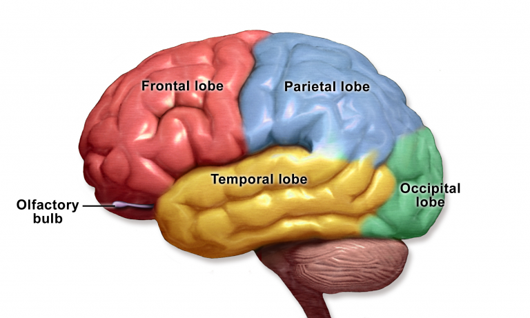
The Four Lobes of the Cerebrum
Each hemisphere of the cerebrum is divided into four lobes: the frontal, parietal, temporal, and occipital lobes. These lobes are defined by regions of the cerebral cortex, which is the outer layer of the cerebrum. It is important to remember that the brain is always communicating across regions. While different lobes have specialized functions, they do not work in isolation.
Each lobe is responsible for specific functions, such as executive control, sensation, memory, and vision. Together, the cortex across all lobes is responsible for the higher-level processes of the human brain, including reasoning, language, decision-making, and personality. Understanding lobe functions helps social workers make sense of client symptoms. For example, frontal lobe damage can affect impulse control, parietal lobe injury may affect sensation and spatial awareness, temporal lobe dysfunction may influence memory or language, and occipital lobe injury may impact vision. Recognizing these links allows social workers to adapt interventions to client needs.
The Brain Is Interconnected
Remember: While different lobes have specialized functions, they do not work in isolation. For example, remembering a story may involve the temporal lobe for storing the memory, the frontal lobe for organizing thoughts, and even the occipital lobe for recalling visual details.
Note: Click on a heading to expand the information.
The Limbic System
The limbic system consists of the hypothalamus, thalamus, the hippocampus and amygdala. The structures and interacting areas of the limbic system are involved in motivation, emotion, learning, and memory.
Emotions: Limbic System Khan Academy Video
Thalamus and Hypothalamus
| Feature | Hypothalamus | Thalamus |
|---|---|---|
| Location | Just above the brainstem, near the pituitary gland. | Near the hypothalamus, between the cerebral cortex and midbrain. |
| Primary Role | Regulates automatic body functions and hormones. | Acts as the brain’s relay station for sensory and motor signals. |
| Functions | Controls body temperature, hunger, thirst, sleep, circadian rhythms, and emotional responses; directs the pituitary gland. | Relays sensory input (sight, sound, touch) to the cortex; sends motor signals to the spinal cord; regulates alertness and sleep. |
| Social Work Connection | Stress, trauma, or sleep problems may reflect hypothalamic involvement, highlighting the link between biology and emotional well-being. | Clients with focus, sleep, or alertness challenges may have thalamic involvement, affecting how they process and respond to the world. |
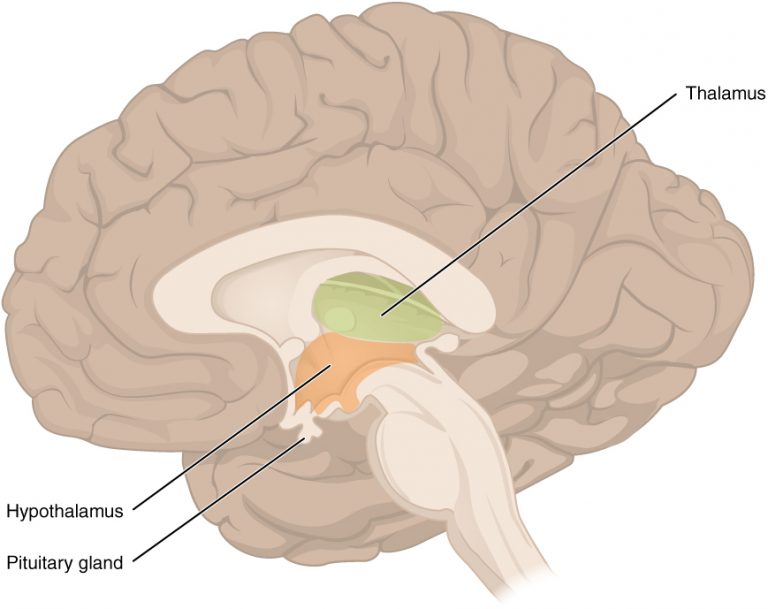
The thalamus, located close to the hypothalamus, acts like the brain’s relay station. It passes sensory information (such as what we see, hear, or feel) to the cerebral cortex for processing, and it also sends motor signals back down to the spinal cord. The thalamus also helps regulate consciousness, sleep, and alertness. Since the thalamus helps regulate awareness and sensory processing, damage or dysfunction can change how a client perceives and responds to the world around them. Difficulties with focus, sleep, or alertness may reflect issues in this area of the brain.
The hypothalamus, a small structure just above the brainstem, plays a big role in keeping the body in balance. It regulates many automatic functions, such as body temperature, heart rate, hunger, thirst, fatigue, sleep, and circadian rhythms. It also serves as an important emotional center, linking the brain and body by responding to signals like light, stress, hormones, and even illness.
One way the hypothalamus influences the body is by producing hormones. For example, it makes oxytocin, which is involved in childbirth, lactation, and bonding, and antidiuretic hormone, which helps the body manage water balance. More importantly, the hypothalamus directs the pituitary gland, sometimes called the “master gland,” to release hormones that control other glands in the endocrine system, shaping stress response, growth, reproduction, and overall health.
Since the hypothalamus regulates stress, emotion, sleep, and hormones, disruptions here can affect both physical and mental health. Clients experiencing trauma, chronic stress, or sleep problems may be showing signs of hypothalamic involvement. Understanding this connection shows how biological stress responses interact with emotional well-being.
“Your brain is always regulating your body. Your body is always sending signals back to the brain.
It’s reporting on the metabolic state of the body…..The brain makes itself aware, not of the little details, but
it makes itself aware of the metabolic status of the body as mood. ” – Dr. Lisa Feldman Barrett
Hippocampus
The hippocampus, located deep in the temporal lobe, is essential for learning and memory. It also helps regulate motivation and emotion, linking experiences to how we feel and act. Because the hippocampus links memory and emotion, trauma can affect how memories are stored and recalled. Clients may struggle with intrusive memories or difficulty distinguishing between past and present danger.
Highlights:
-
The hippocampus, located deep in the temporal lobe, is essential for forming new memories and recalling past ones.
-
It also supports spatial navigation, helping us remember places and directions.
-
This area of the brain is highly sensitive to stress. Chronic trauma or stress can shrink the hippocampus, disrupting memory and learning.
-
The hippocampus is one of the few brain regions where new neurons can form throughout life (neurogenesis), offering hope for recovery and growth.
-
In aging, the hippocampus is one of the first areas affected in Alzheimer’s disease, leading to memory loss.
Amygdala
The amygdala is the part of the brain responsible for formation and storage of memories associated with emotional events. When the amygdala is overactive, clients may appear reactive, anxious, or “stuck” in survival mode. Trauma-informed social work practice can help clients regulate emotions, build safety, and strengthen coping strategies.
| Feature | Key Points |
|---|---|
| Location | Deep within the temporal lobe, close to the hippocampus. |
| Primary Role | Processes emotions, especially fear, stress, and threat detection. |
| Memory Link | Stores memories tied to strong emotions, making emotionally charged experiences easier to recall. |
| Stress Response | Can become overactive after trauma, leading to heightened fear, anxiety, hypervigilance, or exaggerated startle responses. |
| Development | Plays a key role in emotional learning and responses beginning in childhood. |
| Connections | Works with the hippocampus to link emotion and memory, and with the prefrontal cortex to regulate and interpret emotional responses. |
| Impact of Stress/Trauma | Chronic stress can strengthen amygdala activity, keeping clients in “survival mode” and disrupting emotional regulation. |
| Positive Role | Also helps process positive emotions like pleasure and bonding, not only fear. |
Self-Check
