12 The Relationship of the Four Radiographic Properties
This last section of the book deals with radiographic techniques. In this chapter you will learn to analyze the effect of any change in exposure conditions on the four radiographic properties of IR exposure, contrast, spatial resolution and distortion. You will learn how to make changes in exposure factors when one or several exposure conditions change.
Learning Objectives
At the end of this chapter you should be able to:
- Explain the effect on IR exposure, contrast, spatial resolution and distortion with either an increase or decrease in the following factors:
- mA
- exposure time
- mAs
- kVp
- SID
- OID
- focal spot size
- grid ratio
- collimation field size
- filtration
- Calculate mAs, mA or time
- Calculate the new mA and time to be used to control motion, select the small focal spot and use a breathing technique.
- Calculate the new mAs required to compensate for a change in:
- SID
- kVp
- grid ratio
- collimation field size
Throughout this book, you have learned how various factors affect the radiographic properties of IR exposure, contrast, recorded detail and distortion. You have learned the concepts in bits and pieces. This chapter helps you put it all together so that you can see the relationship among the four radiographic properties. A good radiographer must be able to anticipate the effect of changes in exposure conditions on the quality of the radiograph.
This chapter will also review the technique calculations that were each presented separately in other chapters. The math that will be reviewed involves the factors the radiographer uses most often in clinical situations.
IR Exposure
Some factors described in this book have an effect on one radiographic property and some have an effect on more than one property. Depending on the way the factors are changed, some changes cause an increase and some cause a decrease in a particular image quality. Some factors don’t affect a particular property at all. A good way to summarize this is to develop a table for each property in which a plus (+) sign indicates and increase, a minus (-) sign indicates a decrease and a zero (0) indicates no change.
Table 13-1 lists the most important factors that affect IR exposure. In this table, one column summarizes the IR exposure changes seen with an increase in each factor, and another column summarizes the IR exposure changes seen with a decrease in each factor. The + for an increase, – for a decrease, and 0 for no change indicate the factors impact on IR exposure after the factor is increased or decreased. For example, as the mA is increased, IR exposure is increased, and as the mA is decreased, IR exposure is decreased. As the grid ratio is increased, IR exposure is decreased and as the grid ratio is decreased, IR exposure is increased. A short explanation of why the change affects IR exposure is included in the table.
Table 13-1: Evaluation of IR Exposure
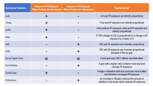
Any factor that affects the amount of radiation produced or transmitted through the patient has to affect IR exposure. mA, time, mAs, kVp and SID all determine the amount of radiation reaching the image receptor. Using any method that controls the amount of scattered radiation that reaches the image receptor also affects IR exposure. These methods include using a large OID in the air-gap technique, as well as the use of grids, collimation and filtration.
Contrast
Contrast will be analyzed in the same way as IR exposure, by developing a table in which one column summarizes the contrast changes seen with an increase in each factor, and another column summarizes the contrast changes seen with a decrease in each factor. See Table 13-2.
Table 13-2: Evaluation of Contrast
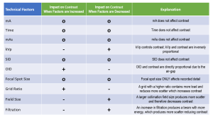
Any factor that changes the amount of scattered radiation produced or the amount of scattered radiation that reaches the image receptor has to change contrast. These factors include using a large OID in the air-gap technique, using a grid, and collimation. Also any factor that changes the penetrating quality of the beam will change contrast. These factors are kVp and filtration.
Spatial Resolution
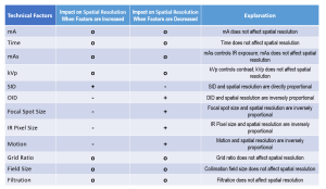
There are only five factors that affect spatial resolution. These factors are SID, OID, focal spot size, pixel size and motion. Table 13-3 lists the effects on spatial resolution, with one column summarizing the spatial resolution changes seen with an increase in each factor, and another column summarizing the spatial resolution changes seen with a decrease in each factor.
Table 13-3: Evaluation of Spatial Resolution
Distortion
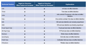
Of the factors listed on the tables for IR exposure, contrast and recorded detail, not many affect distortion. Distance changes always produce more or less magnification, which is size distortion. When magnification decreases, size distortion decreases. A decrease in distortion improves radiographic quality. The factors that affect shape distortion are situational and are difficult to display on a table, so these factors are not included on the tables. These factors are alignment of the central ray, object and image receptor.
Table 13-4 lists the effects on distortion, with one column summarizing the distortion changes seen with an increase in each factor, and another column summarizing the distortion changes seen with a decrease in each factor.
Table 13-4: Evaluation of Distortion
Relationships
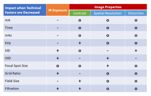
In order to see how the four radiographic properties are related, Table 13-4 lists the effects on all the qualities as the factors are increased. Table 13-5 lists the effects on all the qualities as the factors are decreased.
Table 13-4: Effect on Radiographic Quality if Technical Factors are Increased
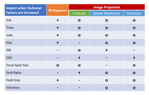
Table 13-5: Effect on Radiographic Quality if Technical Factors are Decreased
Maintaining IR Exposure
Whenever a change is made in an exposure condition, the IR exposure is usually affected. The mAs then needs to be changed to compensate for the effect on IR exposure. This compensation brings the IR exposure back into the satisfactory range. Of course, this assumes that the original IR exposure was satisfactory. The calculations reviewed include: mAs, reciprocity, the exposure maintenance formula, the 15% rule, and grid ratio.
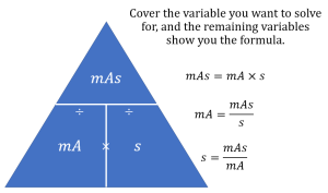
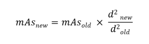
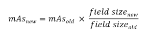
All of the calculations that involve determining a new mAs because of a change in an exposure condition use a similarly structured formula. In this formula, a fraction is formed by placing the multiplier of the new factor in the numerator and the multiplier of the old factor in the denominator. Then the old mAs is multiplied by this fraction and the answer is the new mAs required to maintain the IR exposure after the change in the exposure condition.
milliampere-Seconds
Milliampere-seconds, or mAs, the product of mA and exposure time, determines the quantity of x-rays produced in a beam. mAs, mA and time only affect the IR exposure. IR exposure is directly proportional to changes in mAs, mA or time. This means an increase in mAs, mA or time causes an increase in IR exposure, and a decrease in mAs, mA or time causes a decrease in IR exposure. Exposure time can be stated in fractions, decimals or milliseconds. Milliseconds must be converted to seconds before mAs can be calculated.
Calculating mAs
Reciprocity
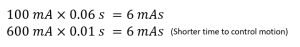
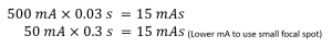
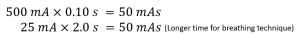
Any combination of mA and time that produces the same product when multiplied will produce the same mAs.
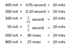
This is the reciprocity law. The radiographer uses this law to control motion by using a short time and therefore a high mA, to select the small focal spot by using a low mA and therefore a long time, and to use a breathing technique by using a long time and therefore a low mA.
Motion Control
Focal Spot Selection
Breathing Technique
Source-Image Distance Changes
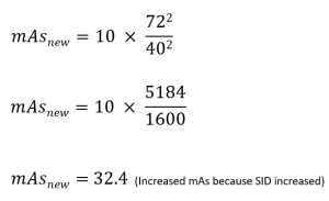
The inverse square law tells us that the intensity (remember this is QUANTITY) of the beam is inversely proportional to the square of the distance from the source. This means that if the source-image distance (SID) is increased, the intensity of radiation at the image receptor decreases, decreasing the IR exposure. And if the SID is decreased, the intensity of radiation at the image receptor increases, decreasing IR exposure.
If the IR exposure was adequate before the distance change, the mAs must be changed after the distance change to maintain the IR exposure level. The change in mAs brings the IR exposure back to the original satisfactory exposure level before the distance change was made. To maintain IR exposure when SID is changed, we apply the Exposure Maintenance Formula, sometimes called the Direct Square Law.
If a radiographer were using 10 mAs at a 40-inch SID and had to change to a 72-inch SID:
Activity: Exposure Maintenance – Distance Changes
Key Takeaways
When using the Exposure Maintenance Formula with distance changes:
- New distance goes on top (numerator)
- Remember to always SQUARE the distance!
Collimation Field Size Changes
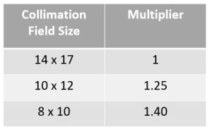
The collimation field size affects the amount of scattered radiation that is produced. A large collimation field size will produce more scattered radiation than a small collimation field size. Since scattered radiation affects IR exposure, a change in the collimation field size will change IR exposure and require a compensating change in mAs if IR exposure is to be maintained. The three commonly used collimation field sizes each have a multiplier that is used in IR exposure maintenance calculations.
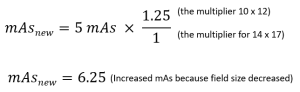
If the original factors used are 5 mAs, 80 kVp with a 14 x 17 inch field size and the collimation is reduced to 10 x 12 inches, what mAs is required to maintain IR exposure?
Activity: Exposure Maintenance – Field Size Changes
Grid Changes
When a grid is used or the grid ratio is changed the goal is to change the amount of scattered radiation on the radiograph and this changes radiographic contrast. Scattered radiation also increases the IR exposure. So if the IR exposure was adequate before the grid change, the mAs must be changed after the grid change to maintain the IR exposure level. The change in mAs brings the IR exposure back to the original satisfactory exposure level before the grid change was made.
To calculate the new mAs to be used in order to maintain IR exposure when the kVp is changed the following multipliers can be used:
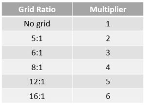
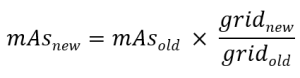
If the original factors are 10 mAs, 75 kVp with a 6:1 grid, and the grid ratio is changed to 12:1, what mAs is required to maintain the IR exposure?
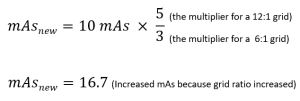
The new mAs produces a radiograph with the same IR exposure as the original, but the contrast will be increased, because the higher grid ratio removes more scatter.
Activity: Exposure Maintenance – Grid Ratio Changes
kilovoltage Changes
mAs is the controlling factor for IR exposure. kVp is the controlling factor for contrast. mAs does not affect contrast; however, kVp does affect IR exposure. If the kVp is increased, more scattered radiation reaches the image receptor and contrast decreases, causing the image to appear grayer (long scale of contrast). An increase in kVp will also increase IR exposure. If the kVp is decreased, less scattered radiation reaches the image receptor and contrast increases, causing the image to appear mostly black and white (short scale of contrast). A decrease in kVp will also decrease IR exposure. An increase or decrease in the kVp of 15% will cause the IR exposure to be doubled or halved. Because mAs is directly proportional to IR exposure, a kVp increase of 15% is equivalent to doubling the mAs. Likewise, a kVp decrease of 15% is equivalent to cutting the mAs in half.
If the IR exposure was adequate before the kVp change, the mAs must be changed after the kVp change to maintain the proper IR exposure. The change in mAs brings the IR exposure back to the original satisfactory exposure level before the kVp change was made. To calculate the new mAs to be used in order to maintain IR exposure when the kVp is changed the following multipliers can be used:
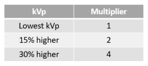
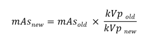
Increasing kVp
If the original factors are 15 mAs and 70 kVp, and the kVp is increased to 80, what mAs is needed to maintain the original IR exposure? Since the original kVp is lowest, we use the multiplier 1 for the kVpold in the numerator. Since the new kVp is 15% higher, we use the multiplier 2 for the kVpnew in the denominator.
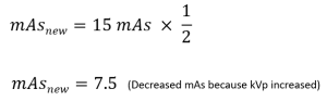
While the new image will show the same IR exposure, it will show lower contrast due to the increase in kVp.
Decreasing kVp
If the original factors are 12 mAs and 80 kVp, and the kVp is decreased to 60, what mAs is required to maintain the IR exposure? Since the new kVp is the lowest, the denominator is 1. Since the original kVp is 30% higher, the numerator is 4.
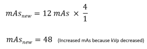
While the new radiograph will have the same IR exposure as the original, the contrast will increase because of the decrease in kVp.
Activity: Exposure Maintenance – kVp Changes
Putting it All Together
Many changes in exposure conditions require a compensating change in mAs to maintain IR exposure at optimum levels. While it is prudent to change only one technical factor at a time, there are times when multiple factors are changed at the same time. Because the Exposure Maintenance Formulas for each technical factor are set up in the same way, we can combine the formulas without causing problems. I like to combine the factors in alphabetical order – distance, field size, grid, kVp. You can remember which factors are in the numerator of each fraction by thinking – New goes in the New-merator – except the krotchity old kVp who wants to be on top! Finally, remember to square the distances!
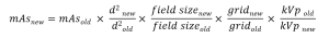
The process for using this combined formula is pretty simple:
- Find the old mAs (if necessary).
- Plug everything into the big Exposure Maintenance formula.
- Square the distances.
- Use the kVp multiplier factors, NOT the actual kVp numbers.
- New goes on top, except for kVp.
- Solve for the new mAs.
- Use the new mAs you just calculated to find mA or time (if necessary).
This formula can be used for any of the exposure maintenance calculations described previously, PLUS can be used to easily calculate the new mAs when multiple factors are changed. If a factor is not changed, the old and new values are the same, resulting in a value of 1. This means you can just ignore any technical factors that do not change because they won’t require a change in mAs to maintain the IR exposure.
This formula can also be used to solve problems where you are given 2 or more complete sets of exposure factors and the question wants to know which set will give you the highest or lowest IR exposure. If you have more than 2 sets of factors, try to narrow them down by looking at the general direction of the factors you are given. Once you are ready to compare 2 sets of factors, assign one set as “old” and the other as “new” and solve for the new mAs just like you did for these questions. Then compare the mAs you got (which is the mAs that is equivalent to the old exposure with the new factors) to the mAs value they provided. If the mAs they provided is higher than your “maintenance” number, that set of factors will give you a higher exposure. If it is lower, then it gives you a lower exposure.
Summary
When an exposure condition changes, one or more of the four radiographic properties may be affected. IR exposure is always affected by a change in the amount of radiation reaching the receptor. Contrast is always affected by changes in the amount of scattered radiation and changes in the beam quality. Spatial resolution is affected only by geometric factors. Size distortion is affected by distance changes. A change in mAs is required to compensate for changes in source-image distance, field size, grid ratio and/or kVp.